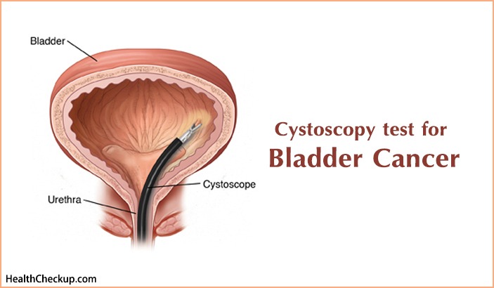
Cystoscopy derives from the thin fiber-optic tube instrument cystoscope that is inserted in your body through bladder to diagnose the internal happenings of your body. A German Army Surgeon Dr. Phillip Bozzinni invented and developed the instrument in 1807. Since then, it has been a significantly remarkable contribution to the urology field of medicine and health. Cystoscope have lens (like telescope or microscope) placed at its inserting end that reproduces the inside image focused upon by enlarging it. Urologist is the specialist Doctor that conducts Cystoscopy test for Bladder Cancer.
Usually stones, blockages, enlarged glands, bleeding, and related bladder abnormalities causes Cystoscopy.
The Cystoscopy test is conducted to find the cause of urinary system problems, bladder cancer, urinary infections, inject a dye for x-rays and kidney problems, collect tissue samples and eliminate foreign objects from the body.
Cystoscopy Procedure:
In some cases, the Urologist Doctor will prescribe antibiotics before and after the Cystoscopy test. You may be asked to vacant your bladder before the Cystoscopy test procedure begins. Anesthetic gel is applied at the urethra tube of your external bladder to numb it as anesthesia. The genitals are treated with antiseptic to cleanse them and a sterile sheet is placed at the area.
The Urologist will insert the Cystoscope tube through your urethra. If the case is of investigative nature of the case, then flexible Cystoscopy test is conducted with a flexible cystoscope. In flexible Cystoscopy test the examinee is instructed to lie flat on the operation bed. Rigid Cystoscopy test serves the operation procedure in Biopsies and surgical cases. In Rigid Cystoscopy test the patient is asked to be situated in a position just as in the pelvic examination. Water or saline is inserted with cystoscope into the bladder that causes the bladder wall to be stretched. This helps the Urologist to examine and investigate the bladder clearly. The Urologist may collect tissue samples for laboratory testing. The inner happenings and functioning of the bladder can be visual on the TV or computer Monitor. Some patients maybe asked for giving urine samples as well before or after Cystoscopy test for bladder cancer.
Usually the Cystoscopy test procedure takes 15 to 30 minutes. With anesthesia the pain is reduced to negligible or very minute that the patient might face during the Cystoscopy test procedure.
Cystoscopy Test for Bladder Cancer:
Bladder cancer goes through four stages. The first stage sees the development of cancer in the inner lining of bladder. In second stage the cancer spreads to bladder wall. In third stage, the cancer cells spread from wall to other tissues. During fourth stage the cancer cells advance to other parts of the body like liver, lungs, etc.
Cystoscopy test for Bladder Cancer provides clear information about condition of problem. There are different procedures for various cases.
Blood Test:
The blood sample is simply collected to determine your general health, the quantity and quality of blood and the functioning of your kidney.
Urine Test:
In this case, the urine sample is examined under the micro-scope to detect cancer cells in a procedure called urine cytology. Usually it is conducted in the early stage.
Cystoscopy:
As mentioned above, a thin fiber-optic tube is inserted into the bladder for internal investigations. This is considered as key test in Bladder Cancer cases.
Transurethral resection of Bladder Tumor (TURBT): In some cases it is also simply termed Biopsy. Here, a cell sample and bladder muscle is collected to test the tumor. It is only conducted when abnormal tissues are found in cystoscopy test results. General anesthesia is provide to the subject in the TURBT process.
Imaging Tests:
These are conducted to determine the structures of your urinary tracts. X-rays, sound waves, magnetic fields, radioactive substance, etc are used to analyze the internal structure of urinary tracts and bladder for further examination. MRI [Magnetic Resonance Imagining] or CT [Computerized Tomography] scans are usually conducted in this procedure. Ultrasound is used to generate sound waves and know the internal structure. A dye is injected into the vein before the procedure.
Bone Scan:
The scanning of bone is executed when any abnormalities are observed in the bones in prior tests. It is to detect if the cancer has invaded to bones.
Usually, subjects don’t experience any side-effects or complications after Cystoscopy test. But some cases may face bleeding problems or difficulties in passing urine. A very rare chance of developing a urinary tract infection might also affect the subject after the Cystoscopy test.
Medically Reviewed By