
The concept of blood-tissue barriers developed in the earlier 20th century. It was noticed that when the dye was injected, they failed to stain the brain and the testes. These findings proposed the concepts of the blood brain barrier and blood testis barrier. Similarly, the concept of blood-retina barrier in the eye was also developed.
On the contrary, another study also found, that the dye was able to penetrate prepubertal testes. Also, the seminiferous tubules were not penetrated by blood vessels and lymphatic vessels. Instead, they were found in the interstitial space between the tubules.
What is A Blood Testis Barrier?
The blood testis barrier (BTB) is a physical barrier which separates blood vessels and lymphatic vessels from seminiferous tubules of testes. It is also called the Sertoli Cell Seminiferous Epithelium Barrier because the barrier is formed between the Sertoli cells of the seminiferous tubules.
This barrier develops only at puberty. It one of the tightest tissue barriers present in the body. This is because it is supported by four different cell junctions. This Sertoli cell barrier or the blood-testis barrier can be compared to the blood-brain barrier.
The blood testis barrier is irreversibly disrupted by heavy metal toxicity like cadmium. Cadmium is found abundantly in industries, refining industries and waste incineration and batteries and is known to cause severe testicular injury.
What are Sertoli Cells?
- The Sertoli cells are a type of sustentacular cell, this means that they provide structural support.
- Follicle Stimulating Hormone (FSH) stimulates the Sertoli cells to secrete androgen-binding proteins into the seminiferous tubules.
- Sertoli cells are of maximum importance when we speak of blood bestis barrier.
- The walls of seminiferous tubules are rich in Sertoli cells.
- Sertoli cells and germ cells produce anti-bacterial and anti-viral substances like interferon and defensins.
In a study conducted on live mice, E.coli was administered into seminiferous tubules.
- It was seen that the bacteria reached a maximum level within 2 days but showed a significant decline by day 5 and complete eradication by 2 months.
- Infiltration of neutrophils (a type of white blood cells) was seen with an increased production of inflammatory cytokines.
- This process was followed by infertility and an irreversible disruption of spermatogenesis.
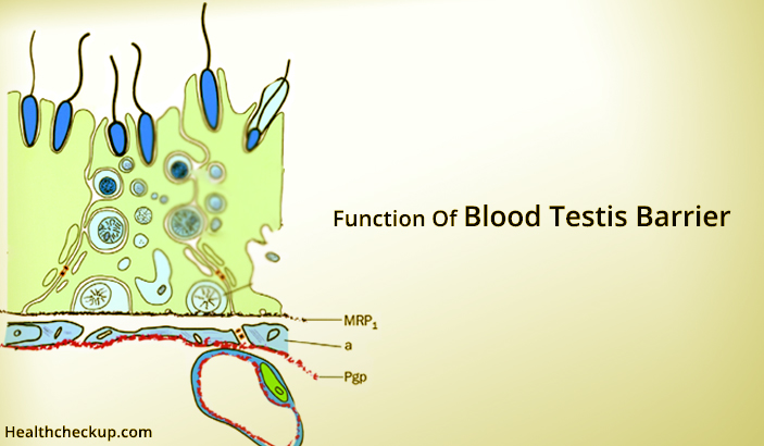
Histology of Blood Testis Barrier
To understand the functions of blood testis barrier, it would be worthy to go through a little bit about its basic anatomical structure and histology.
Blood Testis Barrier is Formed by the Following Structures:
- Tight junction – This is also called the occluding junction. It is a protein complex which provides transport of solutes and ions and also prevents leakage of water and solutes. This function is present only in vertebrates. This tight junction prevents direct contact of spermatozoa with the external environment.
- Adherens junction – This is an intermediate junction, again made of proteins.
- Gap junctions – This is often found in nerves. Gap junctions work as a connection between the cytoplasm of two cells which allows transport of solutes and ions between each other.
- Basal ectoplasmic specializations – These are exclusively present in the testicular seminiferous tubules. This junction is also present between the Sertoli cells. Basal ectoplasmic specialization helps in intracellular adhesion and also plays an important role in spermatogenesis in coordination with Leydig cells.
- Desmosome – This is an intermediate filament based anchoring junction and a flexible adhesive junction.
Studies have found that testosterone and cytokines act together in maintaining the integrity of these junctions between the Sertoli cells. A study also found that Vitamin A was also essential for maintenance of these junctions.
In developing males, that is up to 8 years of age, these junctions between the Sertoli cells are absent.
In patients with hypogonadism, it is found that there is delayed the formation of these junctional complexes.
All these above mentioned types of junctions are present between the Sertoli cells which help in keeping the adluminal environment sterile.
Functions of Blood Testis Barrier:
Blood testis barrier is an essential barrier within the body. Some functions of the blood testis barrier are
- Controls and Maintains the Adluminal Environment : The Adluminal environment is the area near the seminiferous tubules. Germ cells mature into spermatozoa in this environment.
- Controls Passage of Toxic Substances: Like cytotoxic drugs, chemicals and micro-organisms like bacteria, viruses and fungi into the seminiferous tubules. Therefore, maintaining a sterile environment.
- Maintaining Fluid Properties: The blood testis barrier maintains the fluid within the seminiferous tubules. This fluid contains hormones like estrogen and testosterone, inositol (carbohydrate), glutamic and aspartic acid and potassium. Proteins and glucose are present in smaller quantities.
- Maintains Osmotic Pressure: The blood testis barrier maintains a normal osmotic pressure to enable effective transport of solutes and ions from and into the seminiferous tubules. It also controls passage of larger molecules like proteins.
- Antigenic Properties: The germ cell membrane has antigenic properties. The blood testis barrier prevents passage of these antigenic properties into the circulation.
Why is the Blood Testis Barrier Important?
- Provide support – The Sertoli cells provide support to the seminiferous tubules. The Sertoli cells also envelop the germ cells. Under favourable environment, these germ cells differentiate and develop into mature spermatozoa.
- Synthesis – The Sertoli cells synthesize certain substances which are essential for maturation of germ cells.
- Receptors – For Follicle Stimulating Hormones and Testosterone.
- Anti-Mullerian Hormones – Produced by the Sertoli cells. Mullerian ducts in females develop into fallopian tubes. AMH in male fetuses causes regression of the mullerian duct and prevents the development of female secondary sexual characters in them.
- Fertility – Proliferation and mitotic activity of Sertoli cells during childhood and puberty are essential for fertility in adulthood.
- Phagocytosis – Sertoli cells are also responsible for destruction and elimination of degenerated germ cells.
- Spermatogenesis – The blood testis barrier provides a physical barrier to segregate the cellular events of spermatogenesis.
Due to such wide variety of functions, Sertoli cells are also called “Nurse Cells”. The rate of sperm production depends directly upon the amount of Sertoli cells present in males.
Medically Reviewed By

Samarpita is a dedicated freelance writer with avid experience in the space of health, she specializes in topics related to diet, nutrition, immune-related diseases, detection and prevention of diseases and taking a natural route to cure such diseases.