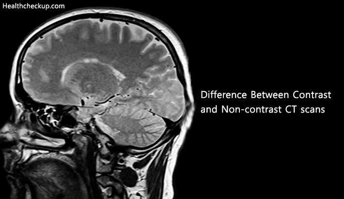
A CT scan also referred to as computed tomography scan, is a medical technique of producing cross-sectional images of particular areas of the scanned part of the body with the help of computer-processed combinations of many x-rays which are recorded from different angles. CT scan was previously known as computerized axial tomography scan or CAT scan. This process helps the medical practitioner to look inside the body part without needing to cut any body part or ask for surgery. CT scan or computed tomography has different forms. Among these forms, two widely known ones are contrast computed tomography scan and non-contrast computed tomography scan.
Background And Statistics
A non-contrast CT scan is a kind of computed tomography scan which is carried out without using a special dye meant to make organs more prominent. CT scans can be done with or without contrast, determined by the medical circumstances of each different case.
A combination of x-rays technology and computer imaging is needed to perform computed tomography scans [CT Scans]. Through this process, horizontal images of the body, also known as slices, are produced. CTs have the ability to provide an interior look into body parts such as organs, tissues, muscles, and bones. A Computed Tomography is one of the most widely used medical tools because of its capability to provide evidence of internal bleeding, lesions, tumors, and many other common health complaints. CT scans can be performed during the patient’s stay at a hospital or in later checkups. Mostly, the chest and abdomen are the subjects of the CT scan process. Both contrast and non-contrast CT scans are regarded as safe procedures having minimal risk and thousands of people worldwide go through these processes every year.
What Does The Procedure Necessitate?
“Dyes” act as contrast agents during a CT scan process. They are mainly concerned with highlighting specific areas to make blood vessels, organs and tissues more visible. With the increasing clarity of all surfaces of tissue or organ undergoing the study, the radiologist can come to know about the intensity and location of injury or disease. Few of the most frequently used contrast agents are:
- Barium
- Gastrografin
- Iodine
- Barium Sulfate
There are a number of ways to administer the chosen contrast agent. These are
- Oral administration
- Intravenous injection
- Rectal administration
- The Inhalation-This method is used in rare cases when a patient is asked to inhale xenon gas for a highly specialized form of lung or brain imaging.
Oral contrast is usually for highlighting the gastrointestinal organs in the pelvis and abdomen. The patient which is to go through oral contrast is directed to fast for a couple of hours before being administered this contrast. There are two types of oral contrast:
- Barium Sulfate and
- Gastrografin.
The former is the most commonly used oral contrast agent. It looks like a milkshake in appearance. The compound is prepared by mixing it with water and it is available in many flavors. Whereas, gastrografin is yellow in color and a water-based drink blended with iodine. When doctors advise oral contrast, patients usually have to drink about 1000 cc to 1500 cc (equal to three to four 12-ounce drinks).
After being swallowed, the contrast reaches the gastrointestinal duct and stomach. X-rays are weakened by gastrografin and barium, as was also the case in intravenous iodine. The organs that have swallowed the contrast look as a highlighted white section on CT image. Normally, both gastrografin and barium contrasts are regarded safe and they ultimately pass through the gastrointestinal tract. Though one may have temporary and minor side effects, like constipation.
When Is Intravenous Contrast Used And Why?
- The purpose of intravenous contrast is to spotlight blood vessels and magnify the structure of different organs such as liver, brain, kidney, and spine. The contrast agent (usually an iodine compound) is clear, with a water-like consistency. This contrast is stored in a special injector which injects the contrast rough a cannula taped in place (often on the backside of hand) during a specific period in the CT exam.
- After being injected into the bloodstream, the contrast circulates in the whole body. The CTs x-ray beam depletes while passing through the organs and vessels that have ingested the contrast. Resultantly, these body parts are emphasized through this process and are exhibited as white areas on CT images. When this test is completed, the contrast is quickly eliminated through kidneys and liver and no harm is caused to the human body. Iodine is regarded as a safe contrasting agent. Medical practitioners have been using this agent for many years without causing any notable side effect on the human body. As iodine provides much visible images of the target tissue, therefore its benefits overshadow the risks it poses.
- The most notable side effect of the said contrast agent is a warm or “flushed” sensation during the process of injection of iodine. Besides this, patients may also feel a metallic taste for one minute in their mouths. If experienced, no treatment is required for this sensation.
- Another benign response may itch over different parts of the body. It may last for several minutes to a couple of hours after being injected. To counter this reaction, medicines are administered to the part of the body where itching is occurring.
- Rectal contrast is employed when to obtain enhanced snapshots of the large intestine and other lower Gastrointestinal tract organs. The contrasts being used for oral contrast are also used for rectal contract, but their concentrations are different.
- Doctors usually use an enema, also known as a clyster, to administer rectal CT contrast. Upon being administered this contrast, the patient may feel a little discomfort, coldness, and a feeling of awash. After the completion of CT, the contrast is eliminated and the patient may visit the bathroom.
- Similar preparations are also needed for rectal contrast as were in oral contrast; patient here too has to fast for many hours before being going through the test. Furthermore, the patient needs to use a Fleets Enema for cleansing the colon; it is mostly done one night before the test.
Differences Between The Contrast And Non-Contrast CT Scans
| Area Of Body | Contrast CT Scan | Non-Contrast CT Scan |
|---|---|---|
| Head CT Scan | Employed for meningeal enhancement, assessment of infection, abscess or tumor for example. Contrast-enhanced head CT is although much inferior to MRI for these indications and very less contrast-enhanced head CTs occur. | Used in case of stroke, headache, trauma and so on. Most things related to neurology use this technique. |
| Chest Computed Tomography (CT) | Employed for testing of the mediastinum (infections and lymph nodes). For evaluating the heart or vascular structures (CT Angiogram for things such as aneurysm, trauma, coronary CT). | Utilized for the purpose of Lung parenchymal evaluation, e.g. interstitial lung disease, lung nodules. |
| Abdomen/Pelvis Computed Tomography (CT) | Essentially everything else, like, Pancreatitis (essential), for the diffusion of abdominal pain, liver mass, abscess, diverticulitis, etc. | Used to find calculi in the ureters or kidneys (the other common sign is acute contrast allergy) Contrast-enhanced scans can make calculi unclear if the contrast begins to get discharged already. |
| CT Angiogram | Utilized for a few indications, like, Haematuria evaluation. Renal masses or abnormal growths in the kidney are found through contrast. | Only employed for a couple of purposes such as Haematuria evaluation. Non-contrast is utilized to find stones. |
What Do You Need To Do Before A CT Scan?
Usually, an appointment letter contains information about everything a patient needs to do before going through the scan, still a few pieces of advice are given that may be useful for a patient.
- You are advised not to eat anything before several hours of your appointment so that clear images can be obtained through the scan.
- In case, you have any allergy or kidney problem, or you are taking medicines for diabetes, you should inform the hospital after having received your appointment letter so that a special arrangement can be made for you.
- It is recommended to wear loose and comfortable clothes so that you do not feel uneasy during the scan.
- Try to go for scan without any kind of jewelry or clothes having metal (like zips), because you will be asked to remove them during the scan.
Where To Get Results?
The results are usually handed over to the medical practitioner that maybe your consultant or primary health doctor. This designated person will then discuss the results and findings with you.
Dr. Adil Maqbool is an M.B.B.S. Doctor and a freelance writer. His ebook on Hipjoint and Sleep has been a well-known success for his clients.