The ophthalmoscopy test is a test performed by an ophthalmologist, a medical doctor who specializes in eye and vision care, to examine the inside of the back of the eye, called the fundus. The fundus consists of the retina, which is a lining of nerve cells on the fundus, and blood vessels and the optic nerve. The retina functions to detect images seen by the outer covering of the eye called the cornea.
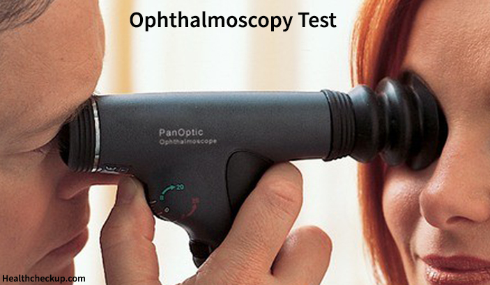
The ophthalmologist uses a magnifying tool called the ophthalmoscope and a light source to see inside your eye. The doctor is able to see other structures in the eye besides the fundus during the ophthalmoscopy test. This test is performed as part of an eye examination or as part of a routine physical examination.
The Two Types of Ophthalmoscopy Test and their Procedure
There are mainly two types of the test, and their procedure is as follows:
- Direct ophthalmoscopy: In this test, you will be asked to be seated in a dark room, and the ophthalmologist will perform the test by shining a bright beam of light through your pupil, using the ophthalmoscope. The ophthalmoscope has the size of a small flashlight, and is made of many magnifying lenses that can magnify up to 15 times, and allow the doctor to view the back of your eyeball.
- Indirect ophthalmoscopy: You will be asked either sit or lie in a semi-reclined position during this test, and the doctor will hold your eye open while shining a bright light into the eye using an instrument worn on his head, that is similar to a miner’s light. The doctor will then view the back of the eye through a lens held close to your eye. He may use a small, blunt probe to apply some pressure to the eye that is being examined, and you will be asked to look in various directions from time to time.
How Each Procedure Feels?
The doctor will use eye drops to dilate your pupils before the ophthalmoscopy procedure. The dilation of the pupils will make it easier for the doctor to see the back of the eye during the ophthalmoscopy procedure. The eye drops will take approximately 15 to 20 minutes to dilate the pupil. Additionally, eye drops may be used to numb the eye surface. The eye drops may cause a stinging sensation in the eye and an unpleasant, medicinal taste in your mouth. You might have trouble focusing your eyes for up to 12 hours after the test, and the tested eye might be very sensitive to light.
During the direct ophthalmoscopy procedure, the light used during the test may feel very strong, and you might see spots for some time after the exam. Sometimes, you may see light spots or branching images, these are in actuality, the outlines of the blood vessels of the retina. Such sensations are absolutely normal.
Indirect ophthalmoscopy might be slightly painful, and the blunt probe used to apply pressure to the eye may cause discomfort. The light used during this procedure is much stronger than that used in the direct ophthalmoscopy procedure, so you might see after-images. You should let the doctor know if you find the procedure very painful.
How to Prepare for the Test?
There are no special steps you need to take in order to prepare for an ophthalmoscopy test, but you should be aware that eye drops will be used to dilate the pupil and/or to numb the surface of your eyes, about 15 to 20 minutes before performing the test. These eye drops may cause you to have trouble focusing your eyes for several hours after the test, so it is always safe to have someone drive you back home, after the test. You will also be required to carry a pair of sunglasses with you to avoid direct sunlight after the test.
Also, it is necessary for the patient to inform the doctor if he/she has glaucoma or if someone else in their family does. You may also inform the doctor if you’re allergic to eye drops, and ask him about the need for the test, how it will be done, and if there are any risks involved.
Why is the Ophthalmoscopy Test Done?
The test is done to help the ophthalmologist:
- Find any diseases of the eye such as retinal detachment
- Spot any changes that indicate a damage to the eye
- Look for any eye changes that may be causing symptoms such as headache
- To detect and evaluate the symptoms of eye diseases such as glaucoma
- To examine the blood vessels of the eye in case the patient has severe hypertension or diabetes and other conditions that affect blood vessels
Risks Involved in the Test
In a few patients, the eye drops used for dilating the pupils or numbing the eye surface before the test may cause symptoms such as nausea, vomiting, dry mouth, flushing or light-headedness. These symptoms should be reported to the doctor immediately. The eye drops might also trigger an allergic reaction or cause closed-angle glaucoma, which is a sudden increase in pressure inside the eyeball. All these symptoms require immediate medical help.
Sometimes patients may experience sudden and severe eye pain or even blindness after the examination. Such patients should call the doctor right away or visit him immediately.
Possible Results of Ophthalmoscopy Test
The ophthalmoscopy test results are said to be normal if all the structures inside the eye including the retina, the blood vessels, and the optic disc appear normal to the ophthalmologist.
However, the results are considered abnormal when:
- There is retinal detachment
- The optic nerve appears to be swollen, causing a condition called papilledema
- The optic nerve is damaged due to glaucoma
- Certain changes such as hard, white deposits appearing beneath the retina, called the drusen or the haemorrhage of the blood vessels of the retina indicate macular degeneration
- Cataracts are found
- Damaged blood vessels or bleeding are spotted in the back of the eye. These damages are usually caused by conditions such as high blood pressure or diabetes.
The ophthalmoscopy test has an accuracy of 95%, and the test is very useful in detecting the early stages of various diseases or their effects on the eyes. The ophthalmologist may carry out other eye tests such as vision tests and tonometry along with ophthalmoscopy to arrive at a conclusive diagnosis.
Medically Reviewed By

I am an experienced Medical/Scientific writer with a passion for helping people live a happy healthy life. My thirst for writing has followed me throughout the years – it is there when I wake up, lingering at the edges of my consciousness during the day, and teases me at night as I go to sleep.