The incidence of coronary artery diseases has risen rapidly in recent years. It is also one of the leading causes of deaths worldwide. Simultaneously, non-invasive techniques in cardiac imaging are being developed which enable early detection and treatment of patients.
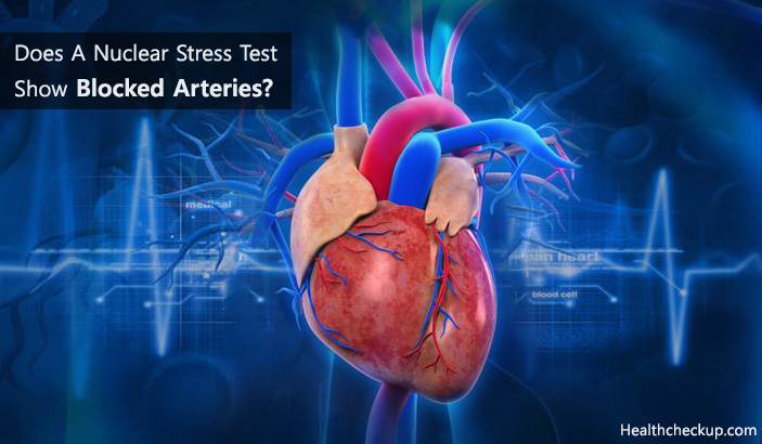
A nuclear stress test is an efficient tool to detect blocked arteries of heart by obtaining images of the heart and its arteries during rest and shortly after exercise.
Does A Nuclear Stress Test Show Blocked Arteries?
A nuclear stress test is used to find out the cause for chest pain brought on by exercise or cause behind unexplained chest pain. A nuclear stress test is used to detect major blockages in arteries of the heart which are responsible for causing coronary heart disease.
A nuclear stress test is done in two phases to compare coronary blood circulation during rest (resting scan) and during exercise (stress scan). Scanning in both phases is done after injecting a radionuclide dye through an intravenous catheter. Areas of blocked arteries will appear as “Defects” or “Cold Spots” because these areas are unable to absorb the dye.
The Purpose Of A Nuclear Stress Test Is To Examine The Following
- A pattern of blood flow within the heart.
- To detect the presence of blocked coronary arteries (blood vessels supplying blood to the heart are called coronary vessels).
- To detect the extent of blocked arteries
- To detect the extent of damage to heart muscles caused by blocked arteries after myocardial infarction (heart attack).
This cardiac imaging tool not only has a diagnostic ability in early detection of blockages of coronary arteries but is also of prognostic value. With the introduction of pharmacological stressor agents, a nuclear stress test can be safely performed in patients who are not eligible to undergo a normal stress test.
This feature enables its applicability in almost all kind of patients. Yet, exposure to radiation remains a potential cause for concern. Newer gamma cameras, however, provide high-quality images with minimum risk of cc.
[Read – Reasons To Fail A Nuclear Stress Test]
How Does A Nuclear Stress Test Show Blocked Arteries?
A nuclear stress test can be performed during a hospital stay or as an out-patient procedure. Before beginning the procedure, the patient is asked to remove all ornaments and change into a dress provided by the scan centre since they may interfere with imaging.
An intravenous catheter is placed on one arm and a blood pressure and pulse monitor on the other arm. Chest leads are applied and connected to an electrocardiogram (ECG). The following two procedures are used to complete a nuclear stress test to detect blocked arteries
1. Nuclear Stress Test with Exercise
- A patient will be asked to walk on a treadmill.
- Intensity, speed, and inclination of the treadmill will be increased gradually.
- Heart rate and blood pressure will be monitored as the exercise continues.
- Once the exercise reaches its peak (at the desired heart rate and blood pressure), a radionuclide dye is injected intravenously.
- After injecting the dye, the patient is supposed to continue walking on the treadmill for a minute or two until the dye circulates.
- The treadmill is then gradually slowed down and stopped.
- The patient is asked to rest for at least 30 minutes before scanning.
2. Pharmacological/Chemical Nuclear Stress Test
- In this phase, the patient is not required to walk on the treadmill.
- A medication or pharmacological agent is injected intravenously to produce stress.
- Heart rate and blood pressure are monitored simultaneously.
- A radionuclide dye is injected through the intravenous catheter.
- The patient will be asked to lie flat and still with arms preferably above the head.
At the end of both procedures, ECG leads are disconnected once the heart rate and blood pressure are again recorded at baseline readings. For scanning, a gamma camera placed above the scanning table takes images of the heart and its arteries in various positions.
It is important to report any unusual signs and symptoms during the procedure such as chest pain, shortness of breath, rash, dizziness or severe fatigue.
What To Expect From Blocked Arteries?
Atherosclerosis is the term used to explain blocked arteries resulting in heart attacks and strokes. This multi-factorial disease is a leading cause for deaths in developed countries and a major burden to the healthcare sector in developing countries.
When the inner lining of medium to large size arteries is injured due to mechanical or environmental factors, inflammatory response develops within the blood vessel.
This leads to an abnormal accumulation of inflammatory cells, lipids and proliferation of smooth muscle cells inside a lumen of medium and large-sized arteries. This lesion is called a Plaque. Progressive development of vascular lesions over a long period of time results in reduced or obstructed blood flow to dependant tissues and muscles.
Certain Risk Factors For Having Blocked Arteries Are
- Age
- Family history
- High cholesterol levels in the blood
- Lack of physical activity
- Cigarette smoking
- Hypertension
- Obesity
- Diabetes mellitus
Patients with atherosclerosis remain asymptomatic for a long period of time. The process of atherosclerosis usually begins during the second decade of life and affects major arteries.
Atherosclerosis can affect arteries of any part of the body. Eventually, signs and symptoms will appear in correlation to the part affected.
Signs And Symptoms For Blocked Arteries In Heart Are
- Pain in left side of the chest.
- Pain radiating to the left arm shoulder or jaw along with profuse sweating is suggestive of myocardial infarction (heart attack).
- Shortness of breath
- Fatigue
Formation of a block within arteries is called thrombosis. Sometimes, a part of this thrombus detaches gets carried by a flow of blood and tends to block smaller blood vessels. This is called embolism.
Other Tests To Detect Blocked Arteries
The prime aim of diagnosis of blocked arteries is to detect their presence and extent before the appearance of clinical signs and symptoms.
- Ultrasonography (USG): This enables observation and evaluation of the degree of plaque formation and narrowing or obstruction to blood flow in peripheral arteries (in arms and legs).
Ultrasonography is also used to detect renal artery stenosis due to atherosclerosis. - Computed Tomography (CT): A CT offers a superior imaging quality as compared to an ultrasound. It enables visualization of coronary arteries after injecting a contrast medium intravenously.
Computed tomography can also help to visualize the extent of calcification and the lipid and fibre content of the plaques. - Magnetic Resonance Imaging (MRI) and Magnetic Resonance Angiography (MRA): This noninvasive test is used to visualize major arteries and stenosis or coronary arteries.
- Angiography: This invasive diagnostic imaging technique is the most commonly used means to detect blocked arteries and their extent by injecting a radio-opaque dye.
- Intravascular Ultrasound (IVUS): It is an invasive form of imaging technique by which arterial wall can be observed by inserting a catheter having an ultrasound probe at its end. The other end is attached to the ultrasound equipment. It enables visualization of plaque volume and properties or content of plaque.
- Angioscopy: It is an invasive diagnostic technique to visualize the interior of arteries by introducing a fibro-optic catheter inside the desired artery. It is frequently used to visualize valves, stents and to estimate the colour and properties of plaque.
Dr. Himanshi is a Homoeopathic consultant and currently working as a lecturer in Post-graduate faculty of Homeopathy, Parul University, Vadodara. Completed BHMS and MD in Homeopathy in January 2018 and also has a clinical experience of about 6 years. Personal interests include reading, spending time with family and traveling.