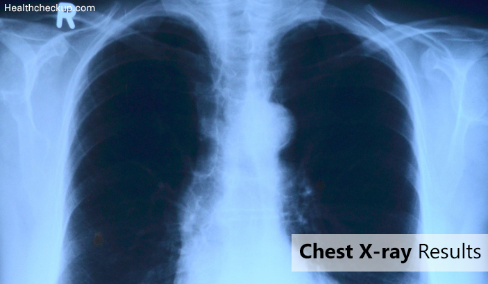
A chest X-ray test is a very common, non-invasive radiology test that provides an image of the chest and the internal organs. Chest x-ray results gives the image of the chest to check if any abnormalities present. First of all, the chest is momentarily exposed to radiation from an X-ray machine and an image is produced on a film or into a digital computer.
The chest cavity captivates different degrees of radiation creating different shadows on the film.
Chest X-ray images are black and white with only the brightness or darkness defining the various structures. For instance, the chest wall bones like ribs and vertebrae absorb more of the radiation and therefore, appear whiter on the film.
The lung tissues are generally filled of air and so allows most of radiation to pass through it creating the darker appearance films. The heart and the aorta will appear less bright like whitish, than the bones.
The Reasons Chest X-ray Tests are Ordered by Physicians are Plenty. But Below are some of the Common Conditions Detected on a Chest X-ray:
- Pneumonia
- Enlarged heart
- Congestive heart failure
- Lung mass
- Rib fractures
- Fluid around the lung (pleural effusion), and
- Air around the lung (pneumothorax).
Overall, a chest X-ray is a simple, quick, inexpensive, and relatively harmless procedure with minimal risk of radiation. It is also widely available.
Why Chest X-ray is Done?
This is Usually Done to:
- Examine the cause of indications like cough, chest pain or shortness of breath.
- Inspect the lung conditions such as lung cancer, pneumonia, chronic obstructive pulmonary disease (COPD), collapsed lung, or if any cystic fibrosis and also to watch treatment for these conditions.
- Find if any heart problems, like heart failure, an enlarged heart, and difficulties causing fluid in the lungs (pulmonary edema), and to monitor treatment for these conditions.
- Check for problems from a chest injury, such as rib fractures or lung damage.
- Find if any foreign objectssuch as coins or other small pieces of metal, in the tube to the stomach (esophagus), the airway, or the lungs.
- See if a tube, catheter, or other medical device has been placed in the proper position in an airway, the heart, blood vessels of the chest, or the stomach.
Chest X-ray Test Procedure
The patients are advised to wear gowns and remove all the jewelleries or any objects or clothes of upper body in order to avoid interference with the tissue visualization. Fasting is not required before chest X-ray as that does not make any difference.
Pregnant women need to notify the doctor and the technician as some or all images may not be taken in order to avoid unnecessary X-ray radiation exposure to the fetus. Precautions, such as, protective lead covers may be placed on the abdomen to avoid radiation to the fetus when an X-ray is absolutely necessary.
Later the following the technician’s advice the patient has to stand in front of a surface nearby the film which records the images. The chest front should be close to the surface. Behind the patient, a machine which releases radiation should be placed. Once the position is appropriate, technician instructs to take a deep breath and hold it and takes the image by device activation. This allows to capture the image on the film in a very few seconds. This leads to development of film in a very few minutes. The film will be sent to doctor for review.
The image can also be taken when the patient is lying provided he/she is unable to stand due to weakness or other medical conditions. This will be called anterior-posterior (AP) view. This is also called portable chest x-ray.
The Conditions and Diseases Recognized by Chest X-ray:
- Congestive heart failure
- Pneumonia
- Emphysema
- Tuberculosis
- Lung mass or lung nodule
- Vertebrae fracture
- Lung fluids
- Rib fractures
- Cardiomegaly
The Risks of Chest X-ray
The risks are very minimal. As X-rays release radiation into the body that is the only slightest risk but it does not last in the body for much time and gets washed once the image has been taken.
Precautions to be taken for pregnant women to minimize the radiation exposure to the fetus.
Chest X-ray Abnormality Detection
Chest X-ray can Detect many Abnormalities which Include:
- Pneumonia
- Lung abscess
- Fluid collection between the lung and the chest wall appearing or pleural effusion
- Pulmonary edema
- Cardiomegaly or enlarged heart size
- Broken ribs or arm bones
- Vertebral fractures;
- Shoulder dislocation
- Lung masses or cancer
- Lung cavities or cavitary lung lesions
- Tuberculosis
- Abnormal presence of air between the chest wall and the lung
- Hiatal hernia
- Aortic aneurysm. These are some of the common abnormal findings that can be seen on chest X-ray Results. There are many other less common abnormalities that can be detected on chest X-ray tests.
Who Interprets a Chest X-ray Test?
Chest X-ray tests are most frequently interpreted by a radiologist (doctor specialized in radiology). Other doctors who often review and interpret the results of chest X-ray tests include internal medicine doctors, pulmonologists, cardiologists, oncologists, etc.
Normally, doctors use the information from a chest X-ray results together with medical history, physical examination, and other clinical data to help to make a clinical decision.
Medically Reviewed By

I am an experienced Medical/Scientific writer with a passion for helping people live a happy healthy life. My thirst for writing has followed me throughout the years – it is there when I wake up, lingering at the edges of my consciousness during the day, and teases me at night as I go to sleep.