
All of us must have heard of Ultrasound test or scan, a very common name in the world of diagnostics. An ultrasound scan, also referred to as a Sonogram, Diagnostic Sonography, and Ultrasonography, is a device that uses high frequency sound waves to create an image of certain parts within the body, such as the stomach, liver, heart, tendons, muscles, joints and blood vessels. Ultrasound scans are non-invasive tests that are useful to detect problems pertaining to the liver, heart, kidney or the abdomen, and they are also useful to surgeons when carrying out certain biopsies.
Although the ultrasound wave travels through the soft tissue and fluids, it bounces back off denser surfaces. The Ultrasound waves will travel through blood, but much of it will echo or bounce back after hitting a heart valve. Similarly, if there are no gallstones in the gallbladder, the ultrasound waves will travel straight through, but when there are stones, ultrasound will bounce back from them.
The bouncing back, or echo, is what gives the ultrasound image its features – varying shades of gray reflect different densities.
Ultrasound is commonly used in many fields of medicine today. Doctors and Health care professionals use sonography for either diagnosis or treatment, as well as for guidance during procedures that require intervention, such as biopsies.
Ultrasounds are used in the field of anesthesiology, cardiology, Emergency medicine, gastroenterology, neonatology, neurology, obstetrics, urology, and also for the musculoskeletal system.
What is Abdominal Ultrasound?
One of the various types of ultrasound scans is the Abdominal ultrasound scan, also known as abdominal sonogram, that is used to check the major organs in the abdominal cavity. These organs include the gallbladder, kidneys, liver, pancreas, and spleen.
Reasons Why an Abdominal Ultrasound is Performed?
The doctor may call for an abdominal ultrasound if there is a suspicion about the possibility of the following:
- Abdominal aortic aneurysms
- Blood clots
- Enlargement of organs such as liver, spleen, or kidneys
- Fluid in the abdominal cavity
- Gallstones
- Pancreatitis
- Kidney blockage
- Kidney stones
- Liver cancer
- Appendicitis
- Tumors
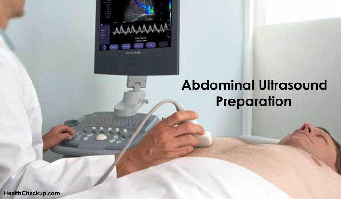
Abdominal Ultrasound Preparation
The Abdominal Ultrasound preparation may entail your doctor to ask you to undergo fasting for 8 to 12 hours before your ultrasound test, as the undigested food can block the sound waves, and make it difficult for the technician to get a clear picture. You should continue to drink water and take your medications as instructed by your doctor.
If you are having your liver, gallbladder, spleen, or pancreas checked, you will need an abdominal ultrasound, and you may need to eat a fat-free meal on the evening before the test, and avoid eating for 8 to 12 hours before the test.
For a test of the kidneys, you may be asked to drink 4 to 6 glasses of liquid about an hour before the test to fill your bladder. You must avoid eating for 8 to 12 hours before the test to avoid gas buildup in the intestines as gas could affect the results of the kidney ultrasound.
If you are having your aorta checked, you may need an abdominal aorta ultrasound, and you have to avoid eating for 8 to 12 hours before the test.
Some people may be worried about what to wear for the ultrasound exam. You should know that you will be asked to remove any clothing, jewelry, and other objects that might interfere with the scan, and you will also be asked to change into a hospital gown. You will be asked to lie down for the ultrasound test. A clear, water-based conducting gel is applied to the skin over the abdomen, which helps in the transmission of the sound waves. A handheld probe called a transducer is then moved over the abdomen. You may need to change positions so that images of different areas can be captured. You may be asked to hold your breath for short periods during the exam.
The ultrasound test doesn’t take more than 30 minutes.
How is Abdominal Ultrasound Performed?
The ultrasound machine creates images of organs and structures inside the body, and sends out high-frequency sound waves that reflect off body structures. A computer receives these waves and uses them to create a picture. The advantage of ultrasound is that unlike x-rays or CT scans, this test does not expose you to ionizing radiation.
What are the Risk Factors for an Abdominal Ultrasound Scan?
An abdominal ultrasound has no risks, whatsoever. Unlike X-rays or CT scans, ultrasounds have no radiation. And, this is precisely the reason why doctors prefer to use them in the case of pregnant women to examine fetuses.
It is possible that you may feel slight discomfort during the procedure, but if you experience pain in your abdomen, bring it to the notice of the technician performing the procedure.
Although there is no concrete evidence of any harm done to fetuses from ultrasound imaging, doctors and researchers are still not sure that there aren’t any long term risks. Ultrasound can heat tissues in the abdomen slightly, and in some cases, it can also produce very small bubbles in certain tissues.
What are the Factors that Affect or Interfere with the Results?
There could be certain factors or conditions that may affect or interfere with the results of the ultrasound test. They are:
- Severe obesity
- Food inside the stomach
- Leftover barium in the intestines from a recent barium procedure
- Excess intestinal gas
What to Expect after the Ultrasound test?
The radiologist will interpret the results of your Abdominal Ultrasound examination. The images would be analyzed and the images sent along with the report to your doctor. Your doctor would discuss with you the results of the ultrasound scan. Depending on what your scan shows, you may be asked to undergo another exam if anything suspicious or questionable was found. If your doctor is able to make a diagnosis of your condition based on your ultrasound, he or she may begin your treatment immediately.
What are the Benefits of Abdominal Ultrasound?
The Ultrasound scan offers many advantages:
- Ultrasounds are painless and do not require needles, injections, or incisions.
- Patients aren’t exposed to ionizing radiation, making the procedure safer than other diagnostic techniques such as X-rays and CT scans.
- The Ultrasound scans capture images of soft tissues that don’t show up well on X-rays.
- Ultrasounds are widely accessible and less expensive than other methods.
Medically Reviewed By