
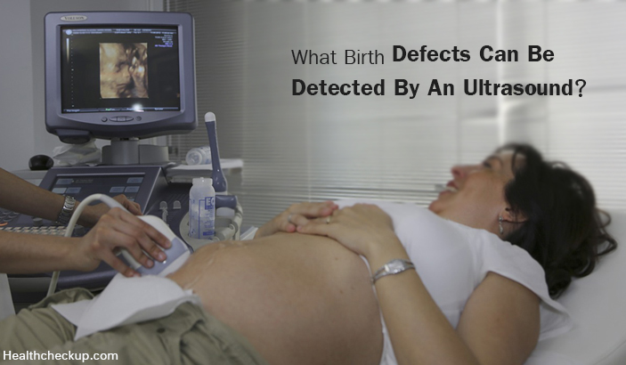
Each pregnancy is associated with a risk of fetal defects and malformations. Screening during pregnancy is essential to understand the health status of a pregnant woman and her fetus. Birth defects have been a major problem worldwide and ultrasound has played an important role in the detection of these. It is a leading cause of perinatal mortality.
In India, the burden of birth defects is significant owing to the large magnitude of the population, poor socio economic status and illiteracy. Additionally, late diagnosis and lack of infrastructure can be blamed for such a high rate of birth defects.
- Annually, about 7.9 million children are born with some serious birth defect worldwide.
- Prevalence of birth defects is 64%. Out of this only 2-2.5% of the birth defects are serious or life-threatening.
- In India, about 495000 children are born with congenital malformations every year.
- About 21,400 are born with Down’s Syndrome, 390000 are born with a G6PD deficiency, 9000 with thalassemia, 5200 are born with sickle cell anemia and 9760 with amino acid disorders.
- About 25-30% of all congenital malformations are due to chromosomal defects.
- Nutritional deficiencies, maternal infections, exposure to drug, radiation and medications account for 5-10% of all birth defects.
Screening During Pregnancy Becomes More Important For Females at Risk, Those Having
- Diabetes Mellitus, Hypertension, Renal Disease
- Above 35 years of age.
- History of giving birth to a malformed fetus
- The family history of a malformed fetus
- Exposure to radiation
- Drugs, Cigarette Smoker, Alcoholics
- Bleeding early in pregnancy
- Consanguineous marriage
Screening in these females is important because there are no specific signs or symptoms which may tell that the fetus may have congenital defects and usually they have an uneventful pregnancy.
During pregnancy, a series of screening tests are done during each trimester. Ultrasound has emerged as an extremely valuable tool is capable of detecting abnormalities in the growth of the fetus.
Ultrasound Is Done In All Three Trimesters For Following Reasons
- To check viability, that is, if the baby is alive or not
- To know the number of fetuses
- An ultrasound scan can detect neural tube defects
- To check for the baby’s normal growth and development
- To monitor placental function
If any abnormality is found, a high resolution ultrasound or a level II ultrasound is done to check for the possible birth defects. Once an abnormality is found via an ultrasound, chorionic villus sampling and amniocentesis are performed to further ascertain the abnormality.
Certain congenital defects, on the other hand, cannot be detected even after the baby is born. Some signs and symptoms may present later in life which suggests a congenital defect.
|
Ultrasound In 1st trimester
|
Ultrasound In 2nd trimester
|
| Done by about 11-13 weeks of pregnancy
|
Done at about 18-20 weeks of pregnancy
|
| Ultrasound scanning in 1st trimester is called nuchal translucency or nuchal fold scan.
|
An Ultrasound scanning in 2nd trimester is called an anomaly ultrasound.
|
| Ultrasound creates images of the fetus and also assesses the status of amniotic fluid around the baby.
This scan assesses risk of baby to have Down’s syndrome.
Birth defects are suspected when extra fluid is noticed at back of the neck.
|
Ultrasound detects size of the fetus and evaluates birth defects in the baby.
Excessive soft tissue at the back of baby’s neck is suggestive of Down’s syndrome.
|
(Read – How Does A Doctor Test for Pregnancy)
What Birth Defects Can Be Detected By An Ultrasound?
| Sr. No.
|
Birth Defect
|
Features Seen On Ultrasound
|
| 1.
|
Trisomy 21 or Down’s Syndrome
Increasing maternal age is often one of the causes for babies born with this syndrome.
|
● Short length of femur and humerus
● Absent nasal bone ● Mild pyelectasis (dilatation of renal pelvis) ● Echogenic intra-cardiac focus on ultrasound ● Short frontal portion of head or brachycephaly ● Abnormal fetal heart rate ● Echogenic intra-cardiac focus ● Short ear length ● Clinodactyly ● Sandal gap deformity |
| 2.
|
Trisomy 13 or Patau Syndrome
It is a rare birth defect. 1 in 1500 babies is born with Trisomy 13. More than 90% babies born with this syndrome have cardiac defects.
|
● Holoprosencephaly – forebrain fails to develop
● Subnormal decrease in distance between two eyes – hypotelorism ● Midline clefts ● Microphthalmia – abnormally small eyes ● Absence of nose ● Echogenic intra-cardiac focus ● Microcephalus ● Ventriculomegaly ● Polydactyly ● Radial aplasia ● Neural tube defect and anterior abdominal wall abnormalities ● Enlarged echogenic kidneys ● Placental abnormality – partial mole
|
| 3.
|
Triploidy
This is a rare chromosomal abnormality where the baby is born with one extra set of chromosomes
Most of the fetuses suffer a spontaneous abortion.
These fetuses have multiple congenital malformations.
|
Severe intrauterine growth retardation (IUGR) if the extra set of chromosome arises from the mother
Large placenta with a partial mole if the extra set of chromosomes arise from the father ● Early onset intra-uterine growth retardation ● Hypertelorism ● Micrognathia – undersized jaw ● Microphthalmia ● Ventriculomegaly ● Dandy-walker malformation – malformation of the brain ● Holoprosencephaly ● Heart defects ● Renal abnormalities ● Syndactyly of third and fourth digit of hand ● Clubbed feet ● Single umbilical artery ● Oligohydramnios ● Spina bifida |
| 4.
|
Turner’s Syndrome
45X karyotype abnormality
The missing chromosome is paternal
Most fetuses suffer a spontaneous abortion. In 2nd trimester fetuses have severe lymphatic abnormalities.
|
● Cystic hygromas
● Pleural effusion, ascites and generalized edema. ● Nearly half of the fetuses have cardiac abnormalities and about19% have renal abnormalities.
|
| 5.
|
Trisomy 18 or Edward’s Syndrome
It is a rare defect. About 3 out of 10000 live-births have trisomy 18.
Babies with this trisomy generally die in the uterus itself due to multiple structural abnormalities.
Polyhydramnios, intra-uterine growth retardation and abnormal hand posture are suggestive of trisomy 18 during 3rd trimester.
|
● Dandy-Walker malformation
● Spina bifida ● Ventriculomegaly ● Clenched hands with overlapping index fingers ● Pre-axial upper limb reduction ● Strawberry shaped skull can be seen in 2nd trimester ● Rocker bottom feet – feet shape resembling that of a rocking chair ● Umbilical cord cysts ● Choroid plexus cysts ● Clubbed feet ● Single umbilical artery ● Polyhydramnios ● Omphalocele – A defect where the baby’s abdominal organs are outside the body due to a hole in the belly. ● Hydronephrosis – Inability of kidneys to drain urine resulting in swelling of the kidney ● Diaphragmatic Hernia – An abnormal opening in the diaphragm through which abdominal organs move into the thorax |
(Read – Tips During Early Pregnancy To Avoid Miscarriage)
How Early Can Birth Defects Be Detected?
An ultrasound to detect birth defects is generally done at about 18-20 weeks of pregnancy. At this time, a detailed scan can be done to observe the growth and development of the fetus. Ideally, each gravid mother should undergo an ultrasound examination at least once a month. If not feasible, a pregnant woman should have an ultrasound done at 11 and 14 weeks and one scan during the second trimester.
The duration for an ultrasound scan can be about 10-20 minutes. It is a non-invasive, cost-effective and a harmless procedure to undergo. Having a normal ultrasound doesn’t always ensure that your child will not have a chromosomal defect. This is because certain minute birth defects cannot be picked up by an ultrasound.
Yet, major congenital deformities are detected during fetal life. Additionally, 3D and 4D ultrasounds have been of additional use to diagnose functional defects accurately. Ultrasound can detect more 60% birth defects efficiently.
Defects like cleft lip, polydactyly, clubbed food, malformed ears, vertebral and other minor structural abnormalities can be easily picked up by 3D and 4D ultrasound scans.
Trans-vaginal ultrasound is becoming increasingly popular. This scan helps in diagnosing birth defects even during the first and early second trimester. Some women may experience discomfort due to the procedure, therefore, a normal ultrasound scan is preferred.
Diagnosis of congenital malformations early in the pregnancy also provides an option of terminating the pregnancy. Because even though the babies are born, their chances of survival are low. Counselling and discussions with the parents can be done.
Certain defects do not necessarily need termination of pregnancy. They can be managed by either treating the baby within the uterus or after delivery without any delay.
Medically Reviewed By
Dr. Himanshi is a Homoeopathic consultant and currently working as a lecturer in Post-graduate faculty of Homeopathy, Parul University, Vadodara. Completed BHMS and MD in Homeopathy in January 2018 and also has a clinical experience of about 6 years. Personal interests include reading, spending time with family and traveling.